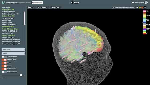
BrainXplore: brain exploration in your browser
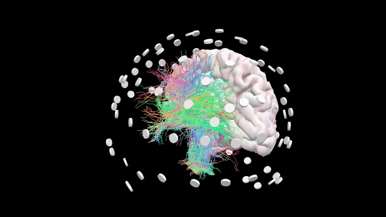
BrainXplore is a browser-based platform for brain exploration that generates patient-specific digital twins from standard structural and functional neuroimaging.
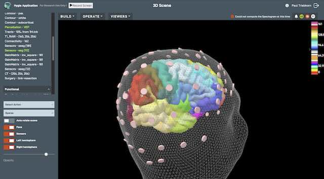
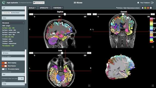
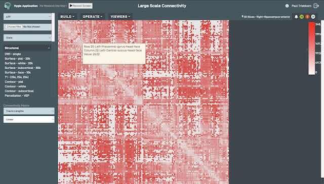
- It provides automated preprocessing, personalized whole-brain simulations that reproduce EEG, MEG, and BOLD dynamics, advanced inference modules, and an interactive 3-D viewer.
- Clinicians and researchers can conduct diagnostics, prototype virtual surgeries or stimulation protocols, and juxtapose simulated signals with each individual’s empirical data.
- Offered for research, education, and investigational clinical studies, BrainXplore streamlines collaboration and accelerates hypothesis testing in neuroscience and precision medicine.
- Built to fit into clinical and research environments, BrainXplore supports standardized data import from multimodal sources and ensures interoperability with existing neuroimaging and electrophysiology systems.